A new study suggests that a short session of gentle electrical stimulation on the ear can quickly shift a brain chemical marker tied to how neurons communicate. The work focused on transcutaneous auricular vagus nerve stimulation, often shortened to taVNS and it used brain imaging to look for immediate changes after a single session.
The study was led by Karina H. Binda and Anne M. Landau at Aarhus University’s Translational Neuropsychiatry Unit. It was published in Psychophysiology. In plain terms, the researchers found that one 30-minute taVNS session in rats reduced a protein signal linked to synapses in several brain areas, while a common “brain energy use” measure stayed about the same.
That mix of results matters because taVNS is often described as a possible tool for brain and mental health conditions. Still, real progress depends on understanding what the stimulation changes in the brain, how fast it happens and what it does not change.
Why Researchers Are Testing Ear-Based Vagus Nerve Stimulation
The vagus nerve is one of the body’s major communication lines between the brain and organs like the heart, lungs and gut. It helps shape things such as heart rate, inflammation and digestion. Because of that reach, scientists have been interested in whether stimulating it can also affect brain circuits tied to mood, thinking and movement.
Traditionally, clinical vagus nerve stimulation has involved an implanted device. That approach can help some patients, such as people with hard-to-treat epilepsy. But surgery is a big barrier. It brings cost, risk and recovery time.
That is where taVNS comes in. With taVNS, stimulation is delivered through the skin at the ear, where a branch of the vagus nerve reaches the surface. In theory, it might offer some similar effects without an operation. Yet the key question remains: what does ear-based stimulation actually do inside the brain, especially right after it is applied?
How The Study Tested taVNS in Rats
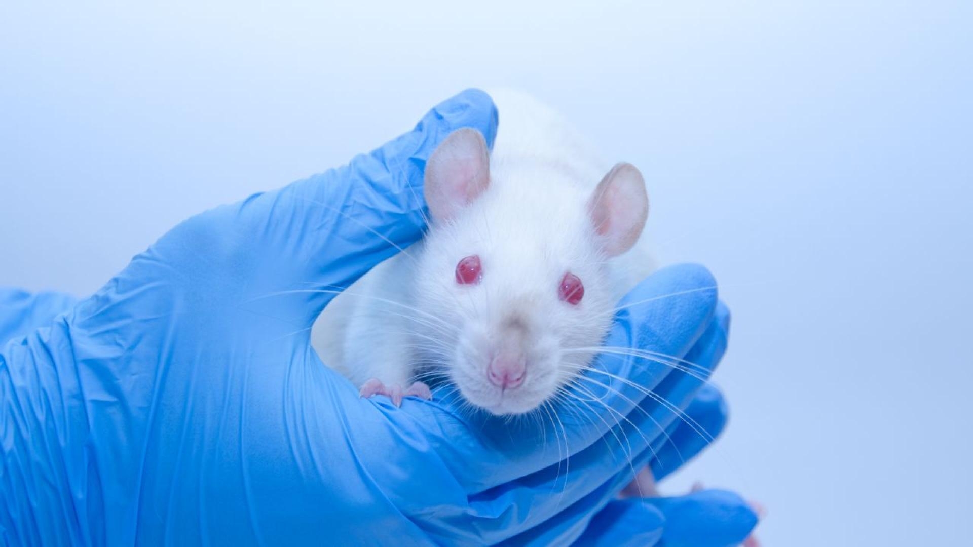
To get closer to mechanism, the researchers used a controlled animal study. They worked with 24 female Sprague – Dawley rats, a common strain in neuroscience research.
One group received real taVNS delivered to the left ear for 30 minutes. Another group received a sham condition, where stimulation was applied to a different body site, the foot, to help control for general sensations and handling.
Importantly, the goal was not to test whether rats behaved differently afterward. Instead, the team aimed to capture immediate biological changes in the brain using imaging methods that can track specific targets.
Two Brain Scans Tracked Synapses and Energy Use
Rather than relying on one measure, the researchers used two different microPET imaging approaches. Each method answers a different question, which helps avoid overinterpreting any single signal.
The first scan used a tracer called [11C]UCB-J. This tracer binds to SV2A, short for synaptic vesicle glycoprotein 2A, which is found in presynaptic nerve terminals. In everyday language, SV2A is closely tied to the machinery neurons use to package and release chemical messages. Researchers often treat it as a synaptic density marker in living brains.
The second scan used [18F]FDG, a glucose-based tracer. FDG PET is widely used as a rough readout of glucose metabolism, which many people think of as overall brain energy use.
Each rat was scanned twice. There was a baseline scan before stimulation, then a second scan afterward. That before-and-after design helped the team spot short-term shifts linked to the stimulation session.
SV2A Levels Dropped After taVNS in Several Brain Regions
The most striking result showed up in the SV2A signal. After taVNS, the rats showed reductions in SV2A binding in multiple brain regions. These included areas often discussed in relation to cognition, movement and mood.
Notably, the reductions were seen in regions such as the frontal cortex, striatum and midbrain. The reported decreases ranged roughly from 36% to 59% across the affected areas. That is a large acute change for an imaging marker, which is one reason this paper stands out.
Another detail made the result even more interesting. Although the stimulation was applied to the left ear, the SV2A reductions appeared on both sides of the brain. In other words, the effect looked bilateral, not limited to the side closest to the ear clip.
Callout: A drop in an SV2A PET signal does not automatically mean synapses were “lost.” In a short window like this, it may reflect quick shifts in synaptic function, neurotransmitter activity, or how the tracer binds. The study adds a clue, not a final answer.
Glucose Metabolism Did Not Change After Stimulation
In contrast to the SV2A findings, the glucose metabolism measure did not show meaningful differences after taVNS. The FDG PET results suggested no broad shift in brain energy use in the immediate period following a single 30-minute session.
That “no change” result matters for interpretation. If taVNS had simply revved up or slowed down overall brain activity, you might expect FDG signals to move. Instead, the pattern looks more selective, at least in the short term.
One way to think about it is that taVNS may nudge certain communication processes without changing the whole brain’s fuel consumption. That does not mean nothing happened. It means the change was not captured by this specific whole-brain metabolic readout, at this time scale.
What The Findings Suggest About Fast Brain Effects
So what could an acute SV2A shift mean? The researchers’ data are consistent with the idea that taVNS can quickly influence presynaptic processes. Those processes include how neurons prepare and release neurotransmitters.
It is tempting to label any change in a “synapse marker” as good or bad. Real biology is rarely that simple. A short-term reduction might reflect temporary dampening in certain circuits, a rebalancing effect, or changes in vesicle cycling. It could also reflect altered tracer binding rather than a physical change in synapse number.
Still, the findings support a key point. taVNS may have rapid effects on brain signaling, even after a single session. That idea fits with why the technique draws interest for conditions where brain circuits may be too “noisy” or poorly regulated.
For readers wondering what this might mean for everyday life, it helps to keep expectations realistic. This study does not show taVNS treats depression or Parkinson’s disease. It does show a possible path for how stimulation might influence brain systems that are relevant to those disorders.
Limits: Small Sample, Female Rats, Single Session
Every study has boundaries and this one has several that are easy to spot. First, the sample was modest, with 24 rats total. Small studies are useful for discovery, but they can be sensitive to chance findings.
Another limitation is that the rats were all female. That was a deliberate and practical choice, but it leaves open a basic question. Would male rats show the same SV2A pattern, or would hormone and physiology differences change the response?
Timing is also critical. This research tested acute taVNS effects after one 30-minute session. Many real-world uses of stimulation, whether implanted or noninvasive, involve repeated sessions over days or weeks. Effects can look very different when the brain adapts over time.
Finally, the study was done in healthy animals. Disease models might respond differently. A brain affected by Parkinson’s-like changes or depression-like changes may have altered neurotransmitter systems before stimulation even begins.
Next Steps: Longer Stimulation and Disease Models
The next stage is to test whether repeated stimulation produces different patterns. Chronic taVNS could lead to changes that are smaller, larger, or even opposite to what was seen after a single session. Longer studies can also check whether imaging changes line up with measurable behavior changes.
Future work can also widen the set of biological markers. If taVNS affects neurotransmitters, inflammation signals, or stress-related hormones, those measures could help explain why SV2A moved while FDG did not.
Another promising direction is to test taVNS in preclinical models of disease. The authors noted interest in conditions like Parkinson’s disease and depression, where brain circuit modulation might be helpful. Testing in disease models can show whether the stimulation effect depends on the starting state of the brain.
Readers who want the technical details can find the original study, which reports the microPET methods, the brain regions examined and the statistical approach used.
Callout: Noninvasive stimulation sounds simple, but the brain’s response can be complex. Studies like this help separate hopeful marketing from measurable biology.